Our original concept of the SC with a “basket weave” appearance at the histological level and a stratum compactum-stratum disjunctum at the electron microscope level has come under scrutiny over the last decade. For instance, Pfeiffer et al. (9) developed new high – pressure freezing followed by freeze substitution techniques for electron microscopy methods and visualized an SC that appeared more compact with smaller intercellular
|
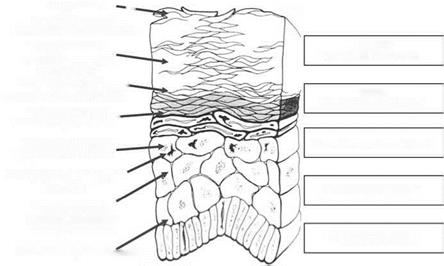
Progressive — corneodesmosomal degradation & desquamation |
|
OUTER STRATUM CORNEUM |
|
T ransglutaminase mediated comeocyte strengthening Dehydration triggers — conversion of filaggrin to NMF |
|
INNER STRATUM CORNEUM |
|
Acidification & lipid „ lamellar bilayer formation |
|
STRATUM GRANULOSUM |
|
Lamellar granule and |
|
NMF precursor-profilaggrin |
|
STRATUM SPINOSUM |
|
T ransglutaminase mediated comeocyte envelope formation |
|
STRATUM BASALE |
|
Cells linked by desmosomes |
|
Figure 2 Typical structure of the epidermis and critical steps in formation of the stratum corneum. Source: From Refs. 1, 3. |

|
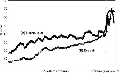
Figure 3 Water profile averaged over a single rectangular region of a cryosection obtained from an individual with (A) good skin, grade 0.5. The horizontal axis is distance across the SC with the SC/granulosum junction indicated by a vertical line. Water profile averaged over a single rectangular region of a cryosection obtained from an individual with (B) dry skin, grade 4. Source: From Ref. 7. |
spaces and hence tighter cell-cell interactions. More controversial, however, was the lack of presence of keratohyalin granules in the epidermis. Lars Norlen (10) has also developed novel cryo-transmission electron microscopy techniques to image vitreous sections of skin without the use of cryo-protectants and, again, more densely-packed SC cells were apparent compared with conventional images and new organelles or tubular structures were observed in the epidermis. Norlen (11) has further proposed a cubic rod packing model for SC keratin structures. However, even with a more compacted SC, several SC swelling regions have been established by Bouwstra et al. (12) and Richter et al. (13) upon skin hydration which appear to be related to loss of barrier function and loss of NMF in the outer layers of the SC, hydrolysis of filaggrin to NMF, and lysis of non-peripheral corneodesmosomes, allowing greater intercorneocyte freedom and transglutaminase – mediated maturation of corneocytes towards the surface layers of the SC. As will be discussed, all of these events become aberrant in dry skin.


