The differences in SC water concentration profiles between normal and dry skin influence the enzymic reactions in the SC. In dry flaky skin conditions, corneodesmosomes are not
|
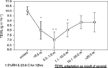
Externally approximately months conditioning 78%RH and 24 C Figure 15 Time course of TEWL adaptation in humans working in an ultra-low humidity environment (1.5%RH). Source: From Ref. 67. |
degraded efficiently and corneocytes accumulate on the skin’s surface layer leading to scaling and flaking. Increased levels of corneodesmosomes in soap-induced dry skin were first reported by Rawlings et al. (23) but have been confirmed more recently by Simon et al. (72). Many corneodesmosomal proteins are now also reported to be increased in the surface layers of xerotic skin. Increased SC corneodesmosomal proteins have also been reported (23,71-73). Interestingly, however, in winter xerosis, the accumulation of the corneodemosomal proteins, Dsg 1 and plakoglobin, correlate with each. Cdsn protein levels, which were also increased, do not, however, have such an association, suggesting that different proteolytic mechanisms occur for the different corneodesmosomal components during desquamation. As suggested by Simon et al. (72), as plakoglobin is a cytoplasmic protein, this would indicate that at least the cytoplasmic domain of Dsg 1 may be cleaved. In fact, immunoreactivity to the carboxy terminal tail of the cytoplasmic portion of Dsg 1 was observed. Perhaps the intracellular portions of Dsg 1 are also degraded within the corneocyte (for example, plakoglobin by the trypsin-like activity or cathepsin E activity reported within the corneocyte matrix). Conversely, Cdsn might be degraded by SCCE, SCTE, or cathepsin D in the lamellar matrix. This is consistent with the early electron microscope images of Rawlings et al. (23) showing that corneodesmosomes become internally vacuolated, followed by complete detachment of the protein structures from the CE (Fig. 8).
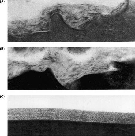
The lamellar lipid matrix is also perturbed dramatically in dry skin (Fig. 16) (23). As the main desquamatory enzymes are found within this lipid matrix, the physical properties of the lamellar lipids will, therefore, influence enzyme activity.
Rawlings et al. (5) originally reported that SC SCCE levels were reduced in the outer layers of xerotic SC compared with normal skin. This has been confirmed recently in more extensive studies by Van Overloop et al. (74) who also found that the equally important SC SCTE activities were also reduced. Conversely, in SLS-induced dry skin, increased activities of these enzymes were reported (28). More recently, the over-activation of the plasminogen cascade has been associated with dry skin. Normally, only observed in the epidermal basal layers, skin plasmin is widely distributed through the epidermis in dry skin. Interestingly, a urokinase-type plasminogen activator also exists in the SC (75). Clearly these and other enzymes are potentially involved in the inflammatory and hyperproliferative aspects of dry skin.
It has been well established that, in hyperproliferative disorders such as dry skin, there is a change in SC lipid composition. In particular, the composition of the ceramide subtypes change and a predominance of sphingosine-containing ceramides (at the expense of the phytosphingosine-containing ceramides) has been observed in the SC of subjects with dry skin. Fulmer and Kramer (76) first identified these changes in SDS – induced dry skin (increased levels of ceramide 2 and 4, and reduced levels of ceramide 3). However, Saint-Leger et al. (77) could not find any changes in ceramide levels in dry skin, but found increased fatty acid levels. Rawlings et al. demonstrated the reduced levels of ceramides at the surface of the SC in winter xerosis (23). At this time, the full complexity of the different ceramide structure was not known, but, more recently, Chopart et al. (78) observed dramatic reductions in the levels of phytosphingosine – containing ceramides in dry skin (approximately 50%), together with a shortening and lengthening of the acyl sphingoid bases sphingosine and 6-hydroxysphingosine, respectively. Van Overloop et al. (74) also clearly demonstrated that the phyto – sphingosine-containing ceramides were reduced to a greater extent than other ceramides, with increasing dryness levels. Fulmer and Kramer at P&G also observed dramatic reductions in the levels of long chain fatty acids in dry skin (76). Imokawa et al. (79) did
|
Figure 16 Organization of stratum corneum lipids in tape stripping of subjects with winter xerosis. Transmission electron micrographs of tape strippings of individuals with severe xerosis. Perturbation in lipid organization towards the surface of the stratum corneum. (A) First strip; disorganized lipid lamellae. (B) Second strip; disorganized lipid lamellae. (C) Third strip; normal lipid lamellae (x200,000). Source: From Ref. 23. |
not find reduced ceramide levels in xerotic skin (but only average levels, rather than superficial levels, were measured).
These changes in lipid composition will, of course, influence the lamellar packing of the lipids. In fact, Schreiner et al. (80) established a reduction of CER EOS and EOH with increased concentrations of sphingosine-containing ceramides (CER NS and CER AS) and crystalline cholesterol in association with a loss of the LPP. However, although the lipid ultrastructure is clearly aberrant in the outer layers of dry skin (23), more work is needed to ascribe a particular lipid phase.
The proportions of the different CE phenotypes also change in subjects with dry skin (43,50). Soap washing leads to a dramatic increase in the levels of the fragile envelope phenotype at the expense of the rigid phenotype (Fig. 17). It is known that SC transglutaminase activities increase towards the surface of the SC, particularly the detergent-soluble and particulate fractions. Although the same trend of the relative increase in TGase between the inner and outer corneum is true of dry skin, TGase activities
Normal □ Soap-Dried
CD r
Q_
 100
100
C
CD
80
ф
60
c
О 40
ъ
20
св
О)
CL
are dramatically lowered in dry skin compared with healthy skin, particularly the detergent-soluble fraction, which contains mainly TGase 1.
Reduced NMF levels are also implicated in dry skin conditions. The loss of NMF generally reported with increased aging, however, is not consistent with the recent observations of Takahashi and Tezuka (81) of increased NMF in subjects with senile xerosis, and suggests that our understanding of this process is far from complete.


