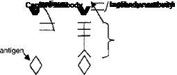
To carry out initial screening experiments, cultured cells are first treated with the potential anti-inflammatory molecule followed by treatment with the inducing agent (ex. IL-1, UVR, TPA), which up-regulates the expression of inflammatory mediators. After a period of time (six to 24 hours), the media is removed and tested for the production of a particular inflammatory mediator using an enzyme-linked immunosorbent assay (ELISA). The ELISA method is based on the recognition of a particular antigen, such as some inflammatory mediator of interest, by a specific antibody, called the capture antibody. While there are many different forms of this assay, one of the simplest variations, the “sandwich assay,” is shown in Figure 6. In this assay, the capture antibody is typically bound to a well in a plastic plate. When the media containing the inflammatory mediator of interest is added to the well, the bound capture antibody binds to the antigen. After binding, the well
is then washed and an additional antibody, called the detection antibody, is added to the well. The detection antibody also binds to the antigen, but in addition this antibody contains a “tag” (for example, an enzyme that reacts with a colorless substrate to produce a colored product) which allows for the amount of bound antigen to be quantified. Thus, if the culture media being tested contains a high amount of the antigen being measured, e. g., IL-1, then a high amount of detection antibody will bind, and a pronounced color reaction will occur when substrate is added. If, however, the anti-inflammatory compound blocks the production of the inflammatory mediator, for example, IL-1, then when the culture medium is added to the assay well, there will be little antigen to bind the capture antibody, and consequently very little detection antibody will bind. The result is very little color formation when the substrate is added.
The advantage of ELISA methods is that they are rapid, can accommodate a large number of samples simultaneously, require very little material for assay (a few microliters of culture medium), are very sensitive (pmole range), and are cost-effective. Commercial ELISA-based assays are available for most cytokines and chemokines, and thus, media from cell cultures can be assayed simultaneously for a variety of inflammatory mediators.
 |
|
Figure 7 shows a flow chart of a screening strategy designed to identify anti – infammatory compounds. As is shown, all putative anti-inflammatory compounds are first screened for the ability to block the IL-1, TPA, or UVR induction of PGE-2, one of the most important inflammatory mediators produced in skin. Although there are exceptions, typically if a candidate anti-inflammatory compound cannot inhibit signaling pathways leading to increased PGE-2 production, it is unlikely to block the production of other inflammatory mediators. Compounds that effectively block PGE-2 production at a concentration of 100 pm or less are then subjected to more demanding dose-response studies and are tested for their ability to block additional inflammatory cytokines and chemokines. For these screening assays, it is important that, where possible, only primary cell strains of human fibroblasts and keratinocytes be used since the use of normal cells increases the probability that results from in vitro studies will be predictive of effects of a given compound when applied topically. Unfortunately, when screening protocols are used for leukocytes, it is difficult to obtain enough normal cells for such studies, and thus, permanent T-lymphocytes and monocyte cell lines are used.
Figure 6 Sequence of steps for enzyme-linked immunosorbent assay.
![]()
![]()
 |
|||||||
|
|
||||||
|
|
|
|
![]()
Gene Array Analysis (5,500 + genes)
Gene Array Analysis (5,500 + genes)
|
|
![]()
Inflammation Specific Northern Assays
Inflammation Specific Northern Assays

![]()
.Candidate Drug
MCP-1 IL-8 IL-12
f /
I LPS-stimulation |
t
THP-1 Monocytes
Figure 7 Screening strategy for assessment of anti-inflammatory activity of a candidate drug.
The results of studies with one putative anti-inflammatory compound are shown in Table 1. The compound was found to effectively inhibit the expression of several inflammatory mediators produced in skin cells in response to various stimuli including UVR, TPA, and IL-1. The table lists the concentration of this particular compound that is effective at inhibiting the induction of an inflammatory mediator by 50% (IC50).
