The CE is an extremely stable and insoluble proteinaceous layered structure. The stability of the envelope is attributed to the degree of cross-linking of envelope proteins by either disulphide, glutamyl-lysine isodipeptide bonds, or glutamyl polyamine cross-linking of glutamine residues of several CE proteins (44). The enzymes, responsible for catalysing the gamma-glutamyl-epsilon-lysine isodipeptide bond formation, are the calcium- dependent transglutaminases (TGase; glutamyl-amine aminotransferases EC 2.3.2.13),
 |
of which four are expressed in the epidermis: TGase 1, 2, 3, and 5. However, only TGase 1, 3, and 5 are thought to be involved in keratinocyte differentiation.
At early time points in the keratinocyte differentiation process, envoplakin and periplakin are expressed and become associated with desmosomes in the viable epidermis. Subsequently, involucrin (the glutamyl-rich protein that covalently-bound lipids become attached to) is expressed at the same time as TGase 1 (45-47). TGase 1 then cross-links involucrin to the other early expressed proteins, such as members of the small proline-rich family of proteins. Subsequently, other plasma membrane proteins become cross-linked, and these form a scaffold for further reinforcement and maturation events (48).
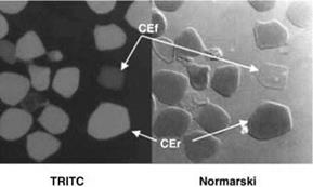
By Normarski microscopy, CEs (CE’s) were shown to have a crumpled surface when isolated from the lower layers of the SC and a smoother, more flattened surface when isolated from the upper SC. These two populations of CEs were named fragile (CEf) and rigid (CEr). Mils et al. (49) reported that about 80% of corneocytes from volar forearm skin were smooth and rigid, whereas 90% from foot sole were rough or fragile cells. They can also be further differentiated by their binding of tetra-methyl rhodamine isothiocyanate (TRITC), with the rigid envelopes staining to a greater extent (Fig. 10) (50). However, Hirao et al. (51) have used a more elegant method to identify CEs based upon their hydrophobicity (staining with Nile red) and antigenicity (to anti-involucrin) (Fig. 11). It is clear from these studies that immature envelopes (CEf) occur in the deeper layers of the SC (involucrin-positive and weak staining to Nile red or TRITC) and that mature envelopes occur in the surface layers of healthy skin (apparent involucrin staining lessened and increased staining with Nile red or TRITC). More recent work from Kashibuchi et al. (52) using atomic force microscopy confirmed these structural changes in corneocytes from the deeper layers of the SC.
The classification of fragile and rigid envelopes has subsequently been found to be a pertinent classification system as, mechanically, they have fragile and rigid characteristics
|
Figure 10 Fluorescence and Normaski phase contrast microscopy of TRITC stained comified envelopes demonstrating increased fluorescence labelling of CEr compared with CEf. Source: From Ref. 50. |
under compressional force (Fig. 12) (50). Supporting this concept of increasing CE strength, gamma glutamyl-lysine cross-links also increase in the subsequent layers of the SC, due to enhanced TGase activity. Three pools of TGase activity have been identified in the SC which have been classified based upon their solubility characteristics: a water – soluble TGase (mainly TGase 1 and 3), a detergent-soluble TGase (TGase 1), and a particulate form that cannot be liberated from the corneocyte. Whether all enzyme fractions are active in this maturation process of CEf to CEr is currently not known.

