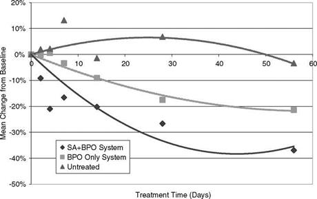
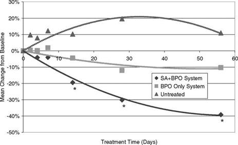
Photography has been a useful tool for evaluating and documenting treatment benefits (Fig. 3 for example). Several acne-grading methods have even been proposed based on photographs. Recent advancement in digital imaging has made image capture and evaluation much more convenient. The techniques that have been applied to clinical imaging go beyond just regular photography that mainly utilizes the visible light spectrum. Photographers have used polarized filters to either cut through the surface specula for a matted appearance with cross-polarized light (Fig. 4 for example), or to enhance the surface specula to make the shine shinier or the surface texture more 3-dimensional-like with the help of parallel-polarized light. Skin redness and pigmentation are highlighted in cross-polarized light images, while parallel-polarized light images bring out the surface luminosity and make the height of a pimple or the depth of a wrinkle more distinctive.
|
|
|
|
|
Baseline 1 Week 8 Weeks
|
Figure 3 Acne skin images from visible light photography comparing the skin conditions before, during and after treatment with an over-the-counter combination therapy system (salicylic acid + benzoyl peroxide). Note the marked improvements in skin texture, bumpiness and clarity. Skin redness was also reduced (data not shown). |
Active
|
Baseline |
Day 4 |
Day 7 |
|
Untreated |
||
|
Baseline |
Day 4 |
Day 7 |
Figure 4 Cross-polarized light images tracking pimple resolution and redness reduction of a pimple treated with a 2% SA gel (active) and of an untreated pimple. Blinded analysis of individual pimple images showed pimple resolution by the 2% SA gel was faster than by the placebo (the vehicle gel) and the untreated control, and similar to that of a 10% benzoyl peroxide lotion. Abbreviation: SA, salicylic acid.
(A)
|
|
(B)
|
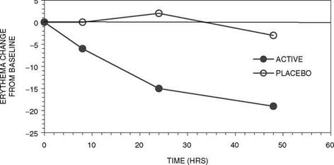
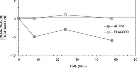
Figure 5 Time Course of target Lesion resolution as measured by hyper-spectral image analysis quantifying (A) Erythema, and (B) Edema (water). |
Another imaging technique involves the use of UV-enriched lamp or blue light. When the skin is illuminated in this manner, the pilosebaceous glands glow as intensely yellow-green or orange-red fluorescent spots (69). Partially or totally clogged pilosebaceous glands all show particularly intense fluorescence of different sizes and brightness, reflecting the degrees to which they are blocked. While the yellow-green fluorescence is associated with the pore plug materials, the orange-red fluorescence is shown to be the emission at wavelengths of 620 and 680 nm by the P. acnes under
385-415 nm light (70-73). The intensity of this orange-red fluorescence is proportional to the density of P. acnes and declines under effective antibiotic treatment (74,75). Fluorescence photography is thus a quick way to assess the antibacterial efficacy of benzoyl peroxide formulations (76).
A powerful method has recently been developed that incorporates all of the above imaging techniques to enable concurrent evaluation of clinical and sub-clinical conditions in the skin (77,78).
The most recent advancement in the clinical digital imaging field is the hyperspectral imaging technique (79). This technique uses narrow-band filters in front of the camera to acquire a series of images (called a hyper-spectral cube). The narrow band filters are selected to detect different chromophores in the skin, the distribution of which is captured in the corresponding images. Each pixel in an image, thus, contains spectral information of the corresponding imaged site on the skin. Reflectance data can be analyzed on a pixel-by-pixel basis to yield chromophore concentrations (oxy-hemoglobin, deoxyhemoglobin, melanin, water, and light scattering). Increased local oxy-hemoglobin concentrations are manifested as erythema. Increased local water concentrations are related to interstitial fluid accumulation due to edema. Chen et al. (80) studied the progression of acne lesion maturation by monitoring lesion erythema and edema with hyperspectral imaging. An example of the results that can be obtained is shown in Fig. 5. Chantalat et al. (81) applied hyperspectral imaging to detect sub-clinical acne lesions that were not yet visible on the skin surface, and tracked the effects of treatments on resolving inflammation associated with the sub-clinical lesions. These studies demonstrate the unique potential of hyperspectral imaging in the evaluation of clinical and sub-clinical acne non-invasively.
Majority of acne sufferers rely upon OTC acne medication to treat their acne. Therefore it is incumbent upon the OTC manufacturers to improve OTC formulations in response to the unmet needs of the average acne patients. While no new OTC acne ingredients have emerged since FDA issued the Final Acne Monograph in 1991, step change has taken place in terms of improved delivery of actives, the choices of vehicles, and the forms of treatments. This is particularly true with daily regimen. It has changed the paradigm of OTC acne therapy from reactive, occasional and irregular usage to routine daily treatment providing the ultimate acne control.
The body of works mentioned in this chapter clearly demonstrates that both benzoyl peroxide and SA are two powerful acne-fighting ingredients. Even though benzoyl peroxide has been the mainstay in OTC acne therapy, our recent work indicates that SA has been under-appreciated. With proper formulation, SA can provide rapid acne clearance with overall efficacy comparable to a 10% benozyl peroxide treatment, while providing high degree of skin compatibility.
In parallel, we have developed powerful, non-invasive imaging and spectral techniques to track acne clinically and sub-clinically at the follicular level. These advanced acne diagnostic methods will enable dermatologists and scientists to develop a much clearer understanding of the acne life cycle in vivo and control its emergence.


