Occipital: The hindmost bone of the skull. Forms the back of the head above the nape.
Parietal: Two bones that form the sides and crown of the cranium.
Frontal: Forms the forehead.
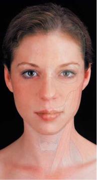
|
FIGURE 2-1: FACE SURFACE ANATOMY Photographer Darrell Peterson remain in his sole copyright. 2005, Elsevier LTD All Rights Reserved |
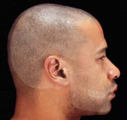
|
FIGURE 2-2: SKULL SURFACE ANATOMY Photographer Darrell Peterson remain in his sole copyright 2005, Elsevier LTD All Rights Reserved |
Temporal: Two bones that form the sides of the head by the ears.
Ethmoid: Between the eye sockets. Forms part of the nasal cavities.
Sphenoid: Joins all the cranium bones together.
Nasal: Two bones that form the bridge of the nose.
Lacrimal: These two bones make up the eye sockets.
Zygomatic (or Malar): The two
cheekbones.
Maxillae: Two bones that form the upper
jaw.
Mandible: The lower jaw, the largest and strongest facial bone.
There are over 600 muscles in the muscular system (Figure 2.4). Muscles are divided into three classes: cardiac, striated (skeletal), and nonstriated (smooth). We deal only with the striated or skeleton muscles that are attached to bones and controlled by will.
Ligaments (which hold the bones together) and tendons (which are connectors between the bones and the muscles) help aid muscles to execute movement. Origin is where the muscle is attached to bones that do not move. Insertion is a muscle attached to a movable bone.


